
EIT Overview
What is Electrical Impedance Tomography?
Electrical Impedance Tomography (EIT) is a measurement technique used in areas as varied as medical imaging and landmine detection [1][2][3]. Electrodes placed on the surface of an object send current and record voltage measurements through the object, from which an internal map of local impedances can be reconstructed. In medical applications, openings and closings of ion channels in neural membranes are reflected as local changes in impedance, as current passing through the object temporarily has more electrical pathways while the channels are open (thus having a measurable lower resistance). Alternatively, regional cerebral blood volume changes as a result of local neural activity, which causes a measurable decrease in cortical impedance. EIT excels over other types of functional imaging as it requires no radioactive tracer, and can reach temporal resolution on the order of milliseconds.
Example EIT Measurement
In the following, we will examine a full measurement cycle of an example EIT system.
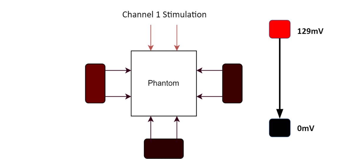
EIT example measurement
One stimulation cycle is shown in the animation above. During each channel's stimulation, a resistor somewhere in the phantom was shorted for 10 ms. The resulting 100 Hz magnitude is represented by a red gradient, where a deeper red corresponds to a larger 100 Hz component in the output voltage for each channel. From this, it is possible to get an intuitive understanding of how EIT works. While Channel 1 stimulates, there is minimal output voltage on any of the other channels, but a small 100 Hz component exists on channel 4. When channel 2 stimulates, there is a larger signal seen, especially on channel 4, but also on channel 3. When channel 3 stimulates, the resultant outputs are similar in magnitude to that of channel 2. However, when channel 4 stimulates, there is a clear increase in the output component at 100 Hz for all channels. As such, we can assume that the resistor that shorted (or the neuron that fired) is far closer to the channel 4 electrodes, where it had the most pronounced effect. In addition, the minimal effect when stimulating through channel 1 also suggests that the resistor was far away from channel 1, likely closer to channel 3.