Faculty Tech Talk: Body Mechanics
Roughly one in ten births in the U.S. is preterm, a higher percentage than almost anywhere else in the world. Prematurity is the number one cause of neonatal death, and those who survive birth face a heightened risk of lifelong disability. Meanwhile, the financial toll on families and the national healthcare system is enormous.
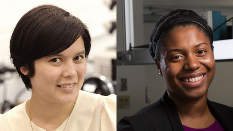
Though disparate factors such as genetics and inflammation are known to play significant roles, root causes of prematurity remain surprisingly poorly understood. By translating insights from tissue mechanics, Columbia Engineering professors Christine Hendon and Kristin Myers are pinpointing how tissue structure itself can contribute to preterm birth—and helping inspire therapeutic interventions that can arrest malfunctions before things go awry.
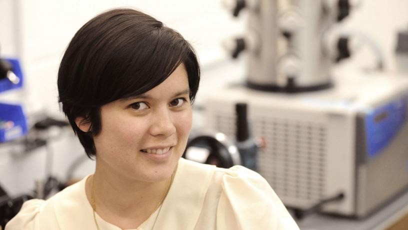
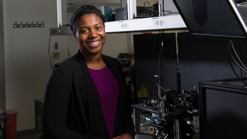
The project serves as a powerful illustration of the deepening intersection between engineering and medicine. Hendon, an associate professor of electrical engineering, develops optical imaging and spectroscopy instruments for scrutinizing the microstructures of tissues such as the heart, joints, and womb. Myers, an associate professor of mechanical engineering, studies the biomechanics of biological soft tissues, particularly in the context of pregnancy and structural mechanisms of birth. They sat down with Dean Mary C. Boyce and a gathering of students for the latest faculty tech talk on November 13, in a dynamic conversation that ranged from tips for effective collaboration to what engineers are uncovering about the structure and function of the human body.
We’ve excerpted a few edited highlights of the conversation below:
Q: Neither of you are biomedical engineers, or at least you didn’t start out that way. What prompted your shift to medicine?
Kristin Myers: I’m a mechanical engineer so usually when I go to a cocktail party or talk to my relatives they’re like, you’re a mechanical engineer, you used to work in automotive – I grew up in Detroit – how did you get into pregnancy? And how do you think of pregnancy as – I don’t want to say a mechanics problem, but an issue that has a lot of mechanics in it.
The structures involved in pregnancy, the uterus and the membranes in the cervix, have these remarkable dual mechanical roles. That’s because in the forty weeks of pregnancy they’re meant to protect the baby. You don’t want the uterus to contract at all, you don’t want the fetal membranes to break, and you don’t want that cervix to open at all. But then at time of labor, they have to completely reverse their mechanical roles. The uterus has to contract, the fetal membranes have to rupture and break, and the cervix has to dilate a lot for a safe delivery of the baby. Those are things that we think of in engineering, fracture and breaking, and they are really important questions for the health of the baby.
Christine Hendon: I really loved electrical engineering as a student and I’m still in the department now because it provides a very nice foundation. My undergraduate major had a biomedical track. I took a course in quantitative physiology in which we modeled body parts with circuits, which was very exciting. In addition, I took a course on biomedical optics that included observing a clinical procedure. It was eye-opening, seeing firsthand some of the current limitations within medicine and knowing that with our traditional engineering training we can develop technologies to address medical needs. I’m constantly incorporating optics and signal processing from traditional electrical engineering to improve our instruments, and I try to incorporate new technologies into our research.
It was eye-opening, seeing firsthand some of the current limitations within medicine and knowing that with our traditional engineering training we can develop technologies to address medical needs.
CHRISTINE HENDON
Q: What recent developments are you most excited about, in your work together and separately?
Myers: In the Soft Tissue Lab we want to build computational models so physicians can visualize the mechanical forces on the uterus and the cervix. We need tools to understand what the cervix is made of, and Professor Hendon’s group is building those tools.
Hopefully one day we can do patient-specific, precision medicine that a clinician could do an ultrasound and pull out her iPhone to compute and understand the loading scenario for that particular woman. We just started recruiting our large cohorts of human patients and doing imaging, and so we have the only datasets in the world that have maternal anatomy plus in vivo cervical mechanical properties. It took a really long time to put it all together, but now I have these troves of data on the mechanics of that interface, which I’m so excited about.
Hendon: The main tool we use in the Structure-Function Imaging Laboratory is called optical coherence tomography (OCT), a real-time imaging method that uses light. It is non-ionizing and very safe. Also, we’re starting to integrate tools from computer vision and animation so that we can start to put our optical images into a three dimensional context. For our collaboration with Dr. Myers, we’re imaging cervical samples with our optical technique. We can see with very high resolution the collagen architecture and orientation, which provides information about the mechanical properties.
Our main application within our lab is in cardiac electrophysiology and to treat cardiac arrhythmias. We’ve been obtaining OCT images of the human heart, and once we put them into anatomical context it was quite eye-opening. We realize now we could actually start to answer interesting questions. Is there a difference between sexes? Are there different patterns with people who have atrial fibrillation versus another cardiovascular disease? We now have the tools to analyze it, and it’s really exciting. We’ve been presenting our optical images to physicians, and they love the visual context we provide. The main treatment for cardiac arrhythmias is ablation, and this procedure currently isn’t very successful. Many patients need to come in for a second surgery, so we want to provide a tool for physicians to reduce this second procedure rate.
Q: You both work very closely with clinicians and medical professionals. How does that back-and-forth shape the research?
Hendon: It’s essential, especially for my work doing translational research where in the end clinicians are going to be our users. In many cases they set the design parameters for our systems. Sometimes we present images that clinicians have never seen before, and we ask them if they had that information how would they use it. Then it becomes a dialogue, and we uncover a lot of things together.
For example, we surveyed physicians at Columbia Medical Center and asked them what’s needed to improve ablation therapy. And overall there are two main responses. One, they really need the real time feedback our imaging provides. And the second is they’re still looking at the underlying mechanisms of atrial fibrillation and how catheter ablation can improve it. That’s a more a basic science question our work can address—how can we understand the underlying substrate and how does that impact the function of the heart?
Myers: Physicians, they’re engineers on the floor; they’re thinking on the fly and they’re problem solving. I feel like through conversations you’re teaching each other, and it’s so miraculous what they know, because they’re hands-on, they’re feeling the patient, they understand the boundary values and everything. I could be doing little mechanics problems in my office, but if I’m not simulating a real-world situation—making computational models that can be used to help patients—then I’m not doing my job. And I find that students that come to my lab really like the back-and-forth. We’re the engineers on the job, we’re on site, we’re the ones solving problems. We see how our tools are actually being used.
— By Jesse Adams
Original article is here.
Hendon Photo Credit: Jeffrey Schifman
Myers Photo Credit: Eileen Barroso